The purpose of this article — to teach ordinary dog owner to determine whether the picture is made of the hip of his dog properly. I will show the correct and incorrect positioning, as well as 2 different sets of X-ray images taken from the same dog in the same day. In the first set of shots positioning was done correctly, a second set of images — incorrectly. You will see that in the case of poor positioning of the hip dog looks worse than it actually is. And you will also see that no matter how to position the dog, you will never be able to turn on the pictures bad hip good.
Radiograph (labeled GOOD Positioning) was made with a 10 month old German Shepherd. While the dog is placed on the x-ray table right positioning of the pelvis quite correct.
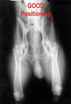
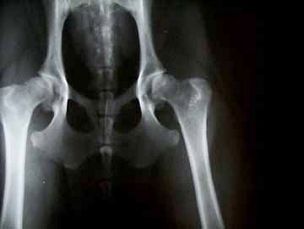
The photo on the right (this is also a photo as above) shows some point shot, which you can check whether the dog is placed correctly.
The first thing that you should pay attention to the picture, whether it lie legs straight down from the hip, knees and straightened if they look alike. We would not like to see that one leg is straightened, and the other comes from an angle.
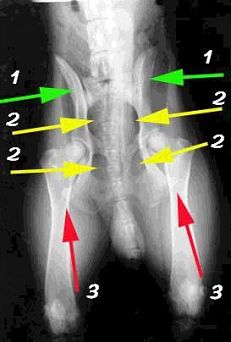
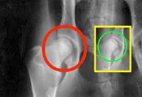
The next shot put three sets of colored arrows (green — 1, yellow — red and 2 — 3).
Green arrows — one point on the bone, in which the recess is a hip joint. These bones look almost like wings. You may notice that the right wing can be seen much better than the left wing. When positioning is 100% true, both wings will be seen almost identically.
Yellow arrows — 2, focused on the gaps in the bone structure. When the correct position of the body, the two lumen of the first side have the same shape and size as the gaps on the right. For a dog in this picture positioning right, but not 100%. Therefore, the left and right gaps are slightly different. It is most noticeable that the lower-right clearance is less than the clearance from the bottom left.
Red arrows — 3 indicate the portion of the hipbone, which is covered with a leg bone in the picture. If you look at the bowl, you can see that with the legs fully extended down the legs overlap both corners or vertices of the pelvic bones. You can see the overlap through the leg bone. In the photo at the top can be seen overlapping equal portions on both sides of the pelvis. In the photo at the bottom can be seen as a much greater overlap in the left part of the photo than on the right. This incorrect positioning. The photo on the right — another radiograph of the same dog. This second picture was taken with incorrect positioning. Notice how much more overlap pelvic bone legs (green arrow — 5) on the left side of the image than on the right. As a result, the bone was pulled out, pulled out away from the trough (red arrow — 4) due to incorrect positioning.
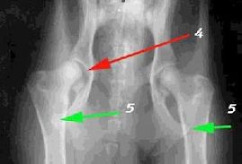
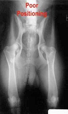
Photo marked POOR Positioning) — example of poor positioning. I repeat that it is the same dog that at the very first, good pictures above. Dog deployed. You can see that the upper right lumen significantly more on the right than on the left. Wing pelvic bone under his feet much more left than the right
The photo below shows the result of poor positioning. In this photo joined two pictures of the same joint of the same dog, taken on the same day. A joint circled round, rounded bone sits deeper in depth than at the joint placed in a rectangle (that happened as a result of improper positioning).
Some people ask how such differences can be striking. I think it’s because we are dealing with young dogs. They still loose ligaments (as in young children). If I had to live through some of the falls, which happened to me at the age of eight, most likely, I would have been a significant number of fractures of the bones. It is the same in dogs. When the dog gets older, her ligaments are not as free and, most likely, can no longer be stretched so much. Such a big difference, as in the photo may not be the older dog, but at a young age is positioned correctly — is critical.
Importance of correct positioning often remains out of focus veterinarians take a picture. There may be several reasons:
Beauceron. buy Beauceron puppy’s lack of experience taking pictures of the hip
Beauceron. buy Beauceron puppy monetary factors. Perform other pictures from the fact that the first wrong, gives them extra money
Beauceron. buy Beauceron puppy during picture position was not correct, because the dog came out of the anesthesia or awake.
In my opinion, among these reasons is no excuse. To get a good x-ray image, you need a good vet. Who started watching the OFA became send pictures back due to poor positioning. When this is done on an ongoing basis, we will have a much better X-ray pictures of our dogs. There are several operations that can be done today to fix a bad hip and allow the dog to live a normal life. The picture to the right — an example might look like the pelvis after surgery. This operation should be done at an early age.
And this picture is very, very bad joints. And it is unlikely that this problem can be solved somehow surgically. This is a snapshot of 8-month-old German Shepherd puppy that was taken from a breeder with the kennel in the backyard. Dog with similar joints should be put to sleep.
The two pictures at the bottom belong to the same dog (Border Collie). The left was made in 8 months of age, the right — at the age of 4 years. Note the thickened neck compound. On a semi-circular portion of traces of arthritis. This dog lives like a home, and for constantly monitoring her condition. When pain occurs, the dog is introduced Rymadil, and I think she feels better.
Ed Frauli, Copyright 1997 (https://www.leerburg.com)